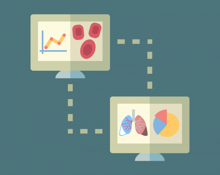
A U-M startup has figured out how to mine and analyze Big Data from digital medical image files such as X-rays, MRIs, and CT scans that could make treatment choices incredibly precise for each patient.
Based in Ann Arbor, Applied Morphomics Inc. uses technology developed at the U-M Medical School to extract thousands of digital biomarkers from a patient’s medical imaging files. From this data, physicians can pinpoint a patient’s condition, the state of their disease and the kind of treatments that might be most helpful.
“This is the ultimate selfie,” said company founder Stewart Wang, the U-M professor whose lab developed the technology. “A patient’s body is their biological medical record and contains a tremendous amount of information that clinicians to date have not been able to comprehend.”